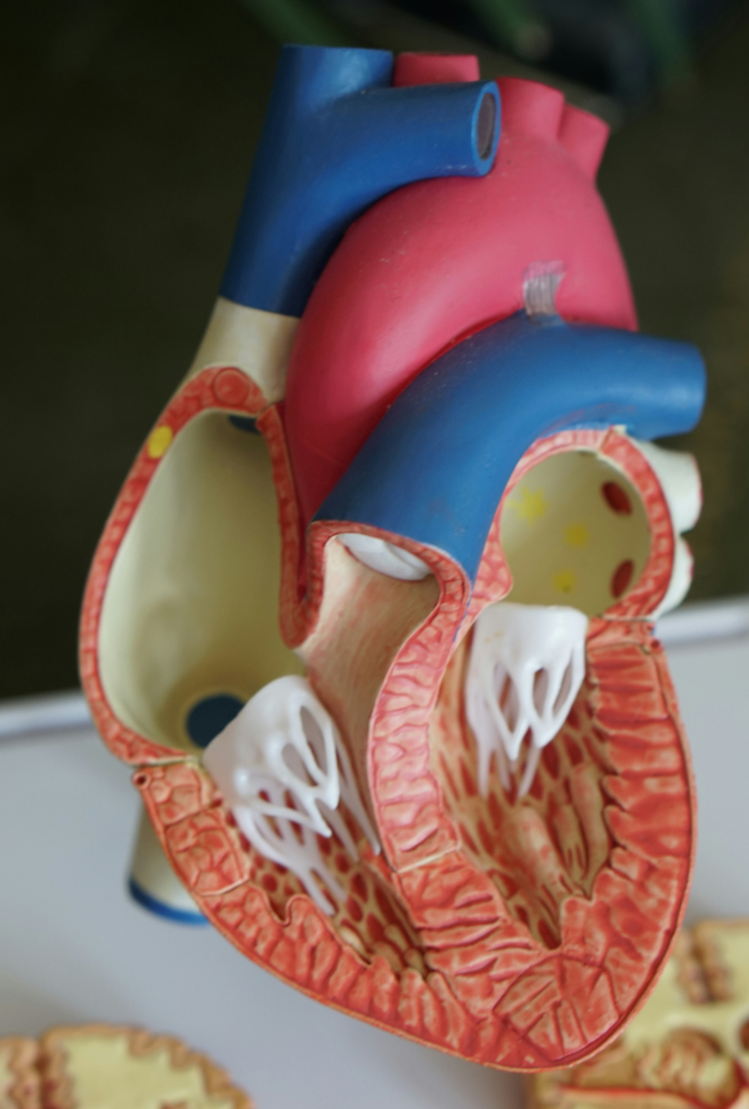
Hypertrophic cardiomyopathy (HCM) is a condition characterized by thickening of the heart muscle (myocardium) that can lead to obstruction of blood flow and increased risk of arrhythmias and sudden cardiac death. Here are the signs and EKG changes associated with HCM:
Signs and Symptoms
- Chest pain: Typically exertional, but can also occur at rest.
- Shortness of breath: Dyspnea on exertion or at rest.
- Fatigue: Reduced exercise tolerance.
- Palpitations: Irregular heartbeats or sensations of skipped beats.
- Lightheadedness: Dizziness or fainting spells.
- Syncope: Fainting, often precipitated by exertion.
EKG Changes
- Left ventricular hypertrophy (LVH): Increased QRS complex voltage (≥35 mm in men, ≥30 mm in women).
- Left atrial enlargement: Prolonged P wave duration (>110 ms) and increased P wave amplitude (>2.5 mm).
- ST-segment depression: Downsloping or horizontal ST-segment depression, often with T wave inversion.
- T wave inversion: Inverted T waves in leads I, II, V4-V6.
- Q waves: Deep Q waves in leads II, III, aVF, V5-V6, indicating septal hypertrophy.
- Pseudoinfarct pattern: ST-segment elevation with Q waves, mimicking a myocardial infarction.
- Bundle branch block: Left bundle branch block (LBBB) or right bundle branch block (RBBB) patterns.
- Atrioventricular (AV) block: First-degree AV block (prolonged PR interval) or higher-degree AV block.
- Ventricular arrhythmias: Premature ventricular contractions (PVCs), ventricular tachycardia (VT), or ventricular fibrillation (VF).
Other Diagnostic Features
- Echocardiography: Thickened left ventricular wall (greater or equal to 15 mm), reduced left ventricular chamber size, and systolic anterior motion (SAM) of the mitral valve.
- Cardiac magnetic resonance (CMR) imaging: Thickened left ventricular wall, reduced left ventricular chamber size, and fibrosis.
- Genetic testing: Identification of mutations in genes encoding sarcomeric proteins (e.g., MYBPC3, MYH7).


Leave a Reply